This is an online elogbook to discuss our patient's deidentified health data shared after taking her/guardian's informed consent.
SHORT CASE :
A 46 yr old female, a labourer by occupation hailing from Nakirekal came to the hospital with Chief complaints of
Shortness of Breath since 5 days and
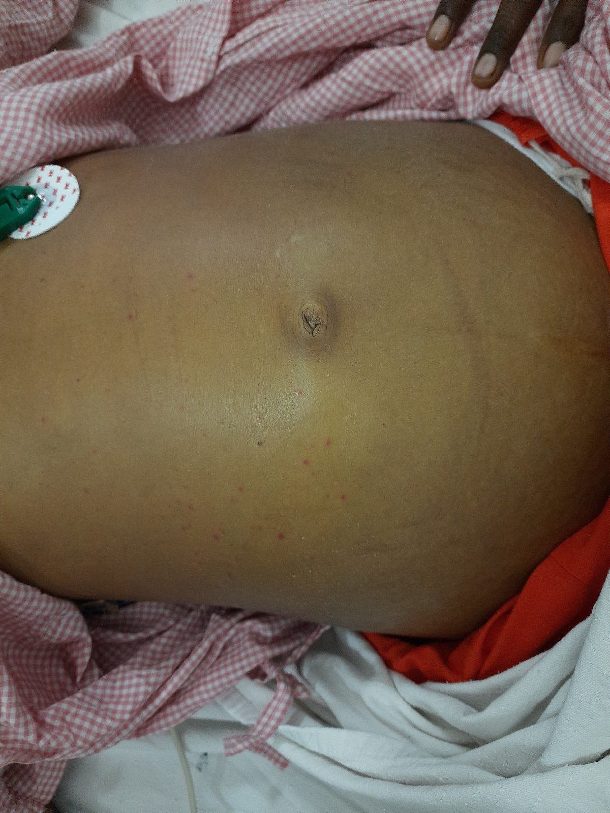
Generalized edema since 5 days.
HOPI :
Patient was apparently asymptomatic 5 days back then she developed Shortness of Breath which was insidious in onset, gradually progressed from Grade 2 to Grade 4 (NYHA grading), aggravated on exposure to dust,cold,lying down and wakes up in the middle of the night and relieved on medication, associated with chest discomfort and Generalized Swelling since 5 days, started in legs first then progressed to entire body.
Associated with cough with mucoid expectoration and wheeze since 5 days.
PAST HISTORY :
Similar complaints in the past every 6-8 months
K/c/o COPD since 8 yrs and on inhalers
K/c/o HTN since 2 yrs and on medication (Telmisartan 80 mg OD)
ON EXAMINATION :
Patient was conscious , coherent and cooperative , well built and well nourished.
Pallor - present
Icterus - absent
Cyanosis- absent
Clubbing - absent
Koilonychia - absent
Lymphadenopathy - absent
Edema of feet - bilateral pitting edema upto the level of knee
Vitals :
Temp -Afebrile
PR- 91 bpm, regular rythm ,voluminous
BP- 110/70 mmHg, right upper arm in sitting position
RR- 28 /min, thoraco abdominal
JVP - raised
Hepatomegaly
Ascites present
CARDIOVASCULAR EXAMINATION:
Inspection -
precordium appears to be normal.
Palpation -
Left parasternal heave
Palpable P2
Apex beat in the left 5th intercostal space lateral to midclavicular line.
Auscultation -
S1, S2 heard
Loud P2
No murmurs
RESPIRATORY SYSTEM :
Inspection - shape and symmetry of chest is normal
Trachea appears to be central
Respiratory movements - rate is increased
Palpation - no tenderness or local rise of temperature
Trachea is central
Apical impulse in the left 5 th intercostal space lateral to midclavicular line.
Increased vocal fremitus is noted in the inframammary areas.
Percussion - dull notes were felt in the infraaxillary and infrascapular areas
Auscultation - bilateral decreased breath sounds and bilateral rhonchi and crepitations present in the inframammary, infraaxillary and infrascapular areas
ECG :
Right axis deviation
Dominant S wave in V5
CXR :Lower lung consolidation
Perihilar hazziness
Enlarged central pulmonary artery
PROVISIONAL DIAGNOSIS :
Right heart failure secondary to COPD (corpulmonale)


Comments
Post a Comment